Science Art Exhibition Eosinophil Portraits
Eosinophil Portraits
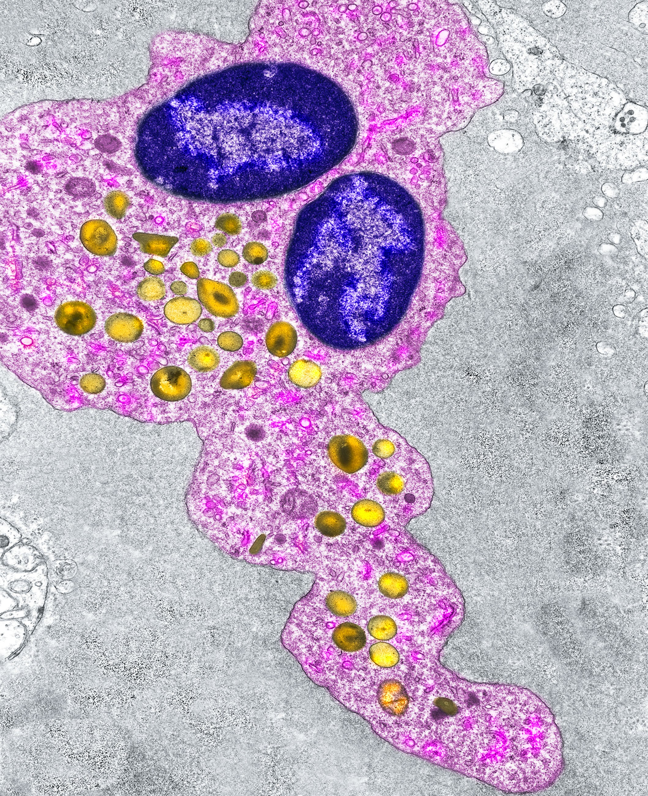 |
This year's meeting featured an art exhibit titled 'Eosinophil Portraits'. Uncovering the unique eosinophil universe, this exhibition features eosinophil images captured by electron microscopy and presented at the intersection of science and art. As a portrait, every image reflects a moment of the eosinophil's lifetime experience in health or disease and invites the viewer to engage with and learn from this multifaceted, enigmatic, and beautiful cell. The exhibition celebrates the release of the book Eosinophil Ultrastructure – Atlas of Eosinophil Cell Biology and Pathology (Elsevier, 2022) by Rossana Melo, Ann Dvorak, and Peter Weller. The book has the distinction of being the first entirely focused on the eosinophil ultrastructure and gathers the studies of three scientists who worked together for decades with a shared passion for the eosinophil world. Photo: A human eosinophil migrating in the small intestine seen under transmission electron microscopy. |